Imaging brain metabolism at the cellular level
Brain metabolism is particularly challenging to study because of the intimate association of multiple cell types (principally neurons and astrocytes) that serve very different functional and metabolic roles in brain tissue.
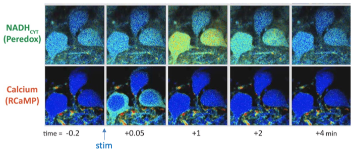
Quantitative imaging of fluorescent biosensors of metabolism, as well as activity detectors such as calcium sensors, can reveal the metabolic behavior of individual neurons or astrocytes in the context of the intact tissue (or even the intact brain). We have used quantitative imaging of the NADH/NAD+ ratio in neurons to assess how their metabolism responds to electrical stimulation (monitored by simultaneous Ca2+ imaging), and to learn how the metabolic responses depend on neuronal glucose metabolism or on imported lactate. We studied both hippocampal dentate granule neurons (in acute brain slice) and layer 2/3 cortical neurons (in vivo). By inhibiting lactate transport or oxidation, we learned that the neuronal metabolic response does not depend on lactate import (e.g. on lactate supplied by astrocytes), but rather on the neurons' own metabolism of glucose via glycolysis.
We continue to study the mechanisms of the fast neuronal metabolic responses to stimulation, using a variety of new biosensors and approaches. Our hope is that improved understanding of neuronal metabolism provides insights into how altered metabolism leads to altered excitability in metabolic seizure resistance, as well as how metabolism may go awry in disease states.